IMRT
IGRT
SRS
Rapidarc
Doctor For IGRT in Delhi NCR, Radiation Treatment in India
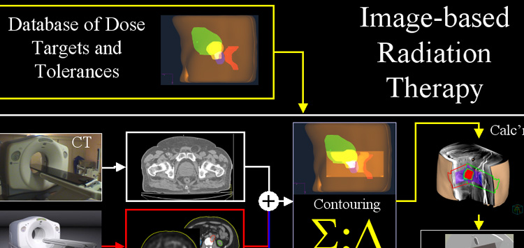
Image-guided radiation therapy (IGRT) is the use of imaging during radiation therapy to improve the precision and accuracy of treatment delivery. IGRT is used to treat tumors in areas of the body that move, such as the lungs. Radiation therapy machines are equipped with imaging technology to allow your doctor to image the tumor before and during treatment. By comparing these images to the reference images taken during simulation, the patient’s position and/or the radiation beams may be adjusted to more precisely target the radiation dose to the tumor. Some IGRT procedures may use fiducial markers, electromagnetic transponders or colored ink tattoos on the skin to help align and target the radiation equipment. If you are to undergo IGRT, your doctor will likely use CT scanning to conduct a treatment simulation session. Other imaging procedures may be used to help determine the exact shape and location of your tumor, and a special device may be created to help you maintain the same exact position during each treatment. Your doctor will give you specific instructions based on the type of exam being performed.

What is Image-Guided Radiation Therapy and how is it used?
-
Image-guided radiation therapy (IGRT)is the use of frequent imaging during a course of radiation therapy for the purpose of improving the precision and accuracy of the delivery of treatment.
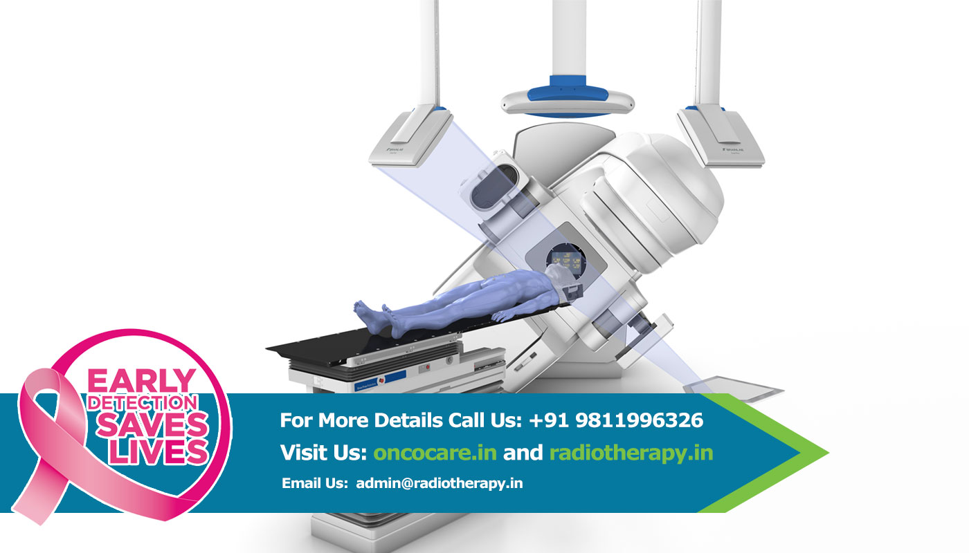
In IGRT, machines that deliver radiation, such as a linear accelerator (for x-ray or photon) or cyclotron/synchrotron (for proton), are equipped with special imaging technology that allow the physician to image the tumor immediately before or even during the time radiation is delivered, while the patient is positioned on the treatment table. Using specialized computer software, these images are then compared to the reference images taken during simulation. Any necessary adjustments are then made to the patient's position and/or radiation beams in order to more precisely target radiation at the tumor and avoid healthy surrounding tissue.
Computed tomography (CT), magnetic resonance imaging (MRI), ultrasound (US) and x-ray imaging may be used for IGRT by visualizing bony or soft-tissue anatomy. Other methods for IGRT use markers placed on the patient's body surface or implanted within the patient's body.
IGRT is used to treat tumors in areas of the body that are prone to movement, such as the lungs (affected by breathing), liver, and prostate gland, as well as tumors located close to critical organs and tissues. It is often used in conjunction with intensity-modulated radiation therapy (IMRT), proton beam therapy, stereotactic radiosurgery, or stereotactic body radiotherapy (SBRT), which are advanced modes of high-precision radiotherapy that utilize computer-controlled x-ray accelerators to deliver precise radiation doses to a malignant tumor or specific areas within the tumor. See the IMRT page, Proton Beam Therapy page or SBRT page for more information.
Who will be involved in this procedure?
-
Delivery of radiation therapy requires a treatment team, including a radiation oncologist, therapeutic medical physicist, dosimetrist and radiation therapists. The radiation oncologist is a physician who evaluates the patient and determines the appropriate therapy or combination of therapies and the type of IGRT. The physician determines what area to treat and what dose to deliver. Together with the therapeutic medical physicist and the dosimetrist, the radiation oncologist determines what techniques to use to deliver the prescribed dose. The physicist and the dosimetrist then make detailed treatment calculations. Radiation therapists are specially trained technologists who acquire images and deliver the daily treatments. The radiation oncology nurse assesses the patient and provides the patient with additional information about the treatment and possible adverse reactions. The radiation oncology nurse also helps manage any reactions or side effects from treatment that may occur, in collaboration with the physician.
What equipment is used?
-
In IGRT, imaging equipment is mounted on or built into the machine that delivers radiation, such as a linear accelerator. Imaging equipment may also be mounted in the treatment room. Imaging technologies used in IGRT include x-rays, computed tomography (CT), magnetic resonance imaging (MRI) and ultrasound (US). Sometimes, IGRT is performed by a detector in the room which tracks motion by localizing markers on the surface of a patient, or electromagnetic transponders placed within the patient.
Who operates the equipment?
The equipment is operated by a radiation therapist, a highly trained technologist. The overall treatment plan is created and supervised by the radiation oncologist, a highly trained physician specializing in treating cancer with radiotherapy.
How is the procedure performed?
At the beginning of each radiation therapy session, the patient is carefully positioned guided by the marks on the skin defining the treatment area. Devices may be used to help the patient maintain the proper position. Images are then taken using imaging equipment that is built into the radiation delivery machine or mounted in the treatment room. The physicians or a radiation therapist then review the images and compare them to the images taken during simulation. The patient may be repositioned and additional imaging may be performed. After any necessary adjustments are made to the treatment plan and patient positioning, radiation therapy is then delivered. The image-guidance process is expected to add additional time to each radiation therapy session.